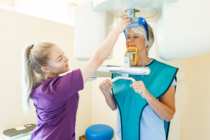
Digital volume tomograph / DVT
Using this special X-ray method, three-dimensional images of the facial skull can be produced in the same quality with a fifth of the radiation dose of a computer tomograph (CT). In addition, our device is able, due to it’s full format image, to display not only a small section, but the entire face skull.
With the help of the 3D image, we can plan an implant on the computer so that the length and width of the implant as well as the direction and depth of the bore can be exactly predetermined. In this way, we ensure the maximum safety of the surrounding structures and a healing process that is as uncomplicated as possible.
The costs of the DVT pictures are usually taken over by the private health insurances. Holders of statutory health insurance are currently required to bear the costs themselves. The costs depend on the type and extent of the DVT examination. We would like to advise you on this subject.